kodymedical@gmail.com
 +91-9380223396
 My Account
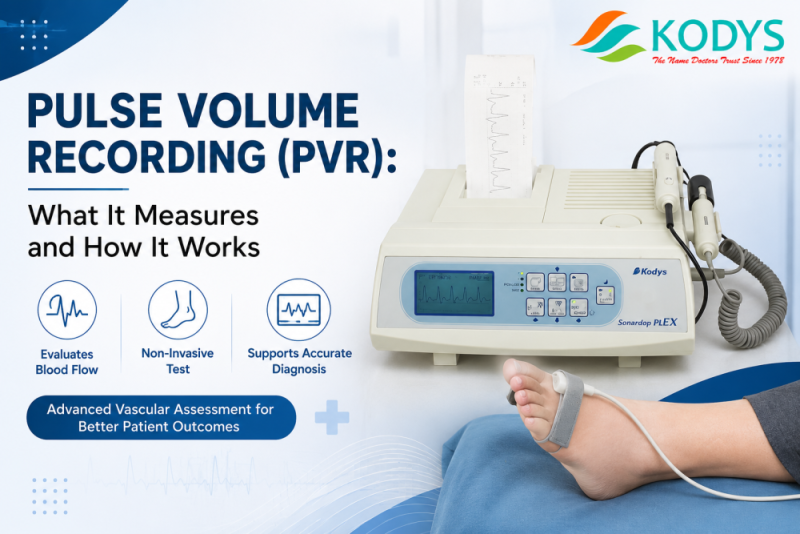
Pulse Volume Recording (PVR) is a non-invasive vascular test that measures changes in limb blood volume with each heartbeat, using cuffs placed on the arms, thighs, calves, ankles, and toes. Kody Medical explains PVR as a waveform-based diagnostic method: a pneumatic cuff detects small volume changes in the limb caused by arterial blood flow, and the resulting waveform shape (triphasic, biphasic, or monophasic) shows whether blood flow through the limb is normal or restricted by peripheral arterial disease (PAD). PVR is typically performed alongside Ankle-Brachial Index (ABI) and Toe-Brachial Index (TBI) testing on automatic vascular testing systems such as the Hadeco SmartDop XT, which Kody Medical supplies to hospitals and diabetic care clinics across India.
What Is Pulse Volume Recording (PVR)?
Pulse Volume Recording (PVR) is a diagnostic test that records the change in blood volume in a limb segment during each cardiac cycle. A blood pressure-style cuff is wrapped around the limb and connected to a plethysmograph, a device that detects small changes in limb circumference as blood flows in with each heartbeat and drains out between beats. These volume changes are plotted as a waveform. In a limb with healthy circulation, the waveform is sharp and triphasic. In a limb affected by arterial narrowing or blockage, the waveform flattens and slows, becoming biphasic or monophasic.
PVR is one of the core components of automatic vascular testing systems used to screen for peripheral arterial disease (PAD), a condition in which arteries supplying the limbs become narrowed, most commonly in patients with diabetes, hypertension, or a history of smoking. Kody Medical supplies PVR as part of its automatic ABI-TBI-PVR vascular testing range, used by diabetic care clinics and hospitals across India to screen for PAD as part of routine diabetic foot assessments.
How Does PVR Testing Work?
PVR testing works by placing pneumatic cuffs at multiple points along a limb, typically the upper thigh, above the knee, calf, ankle, and sometimes the toe, and inflating each cuff to a fixed sub-systolic pressure. As blood flows into the limb segment beneath each cuff with every heartbeat, the limb’s volume increases slightly, and this change is captured by a pressure or air-displacement sensor inside the cuff. The device records this change as a waveform in real time.
The test is performed on both limbs simultaneously so the clinician can compare left and right sides. Because PVR measures volume change rather than direct pressure, it remains accurate even in patients with calcified or non-compressible arteries, a common limitation of standalone ABI testing in long-standing diabetic patients. On the automatic vascular testing systems Kody Medical supplies, this entire cuff sequence runs without manual switching, so a clinician can complete a full multi-segment PVR reading on both limbs in one automated pass.
What Does a PVR Waveform Show?
A PVR waveform shows the pattern of blood flow through the segment of limb being tested, and clinicians classify it into three main types:
- Triphasic waveform: A sharp upstroke followed by a brief reversal and a smaller forward flow. This pattern indicates normal, healthy arterial flow with no significant blockage.
- Biphasic waveform: A blunted upstroke without the reversal phase. This pattern suggests mild to moderate arterial narrowing.
- Monophasic waveform: A slow, rounded upstroke with a delayed peak and no reversal. This pattern indicates significant arterial obstruction and reduced blood flow to the limb segment.
By comparing waveform shape and amplitude at each cuff position (thigh, calf, ankle, toe), a clinician can localize approximately where along the limb a blockage is occurring, which is useful information for planning further vascular evaluation or treatment. Kody Medical’s automatic vascular testing systems display and store these waveforms automatically, so clinicians can review triphasic, biphasic, and monophasic patterns side by side without manual waveform tracing.
When Is PVR Testing Used?
PVR testing is used whenever a clinician needs to evaluate blood flow to the limbs, most commonly in the following situations:
- Diabetic foot screening: to detect early peripheral arterial disease before ulcers or non-healing wounds develop.
- Claudication evaluation: for patients reporting leg pain, cramping, or fatigue during walking.
- Non-compressible artery cases: when a patient’s arteries are calcified (common in long-standing diabetes or chronic kidney disease), making standalone ABI readings unreliable.
- Pre- and post-procedure assessment: to document blood flow before and after a vascular intervention such as angioplasty or bypass surgery.
- Routine vascular risk screening: as part of a combined ABI, TBI, and PVR panel in diabetic and cardiology clinics.
Kody Medical works with diabetic care clinics, vascular labs, and hospital diagnostic departments across India that use PVR as a standard part of their diabetic foot and vascular risk screening protocols.
How Is PVR Different from ABI and TBI?
PVR, ABI (Ankle-Brachial Index), and TBI (Toe-Brachial Index) are complementary tests that are usually performed together on the same automatic vascular testing system, but each measures something different:
- ABI compares blood pressure at the ankle to blood pressure at the arm, producing a single numeric ratio that screens for PAD.
- TBI compares blood pressure at the toe to the arm and is used specifically when ankle arteries are calcified and ABI results would be unreliable.
- PVR does not rely on a pressure ratio at all. Instead, it records the shape of the blood volume waveform at each cuff location, which makes it useful even when calcified vessels distort ABI and TBI readings.
Because each test compensates for a limitation in the others, clinics typically use combined ABI-TBI-PVR systems such as the Hadeco Smartdop XT distributed by Kody Medical rather than relying on any single test in isolation.
What Equipment Is Needed for PVR Testing?
PVR testing requires a plethysmograph-equipped vascular testing system, pneumatic cuffs sized for the thigh, calf, ankle, and toe, and software to capture, display, and store the waveform results. Kody Medical supplies the Hadeco Smartdop XT, a fully automatic vascular testing system that performs ABI, TBI, and PVR in a single sequence and connects to Smart XT Link software for Windows, allowing clinics to generate a combined vascular report for each patient. This system is used across diabetic care clinics, vascular labs, and hospital diagnostic departments in India for structured PAD screening.
Frequently Asked Questions
What does PVR stand for in a medical test?
PVR stands for Pulse Volume Recording. It is a non-invasive vascular test that records blood volume changes in a limb with each heartbeat, using pneumatic cuffs and a plethysmograph. Clinicians use Pulse Volume Recording alongside ABI and TBI testing to screen for peripheral arterial disease, particularly in diabetic patients.
Is a PVR test painful?
No, a PVR test is not painful. The procedure involves placing blood pressure-style cuffs on the thigh, calf, ankle, and toe and briefly inflating each cuff, similar to a standard blood pressure check. Patients may feel mild, temporary cuff pressure during inflation, but pulse volume recording does not involve needles, contrast dye, or radiation.
How long does a PVR test take?
A PVR test typically takes 15 to 30 minutes when performed as part of a combined ABI, TBI, and PVR panel on an automatic vascular testing system. The exact duration depends on how many cuff positions are tested and whether both limbs are assessed simultaneously.
Why is PVR used instead of ABI alone?
PVR is used alongside ABI because ankle-brachial index results can become unreliable in patients with calcified, non-compressible arteries, a common issue in long-standing diabetes. Since Pulse Volume Recording measures waveform shape rather than a pressure ratio, it remains accurate even when arterial calcification affects ABI readings.
Can PVR testing detect diabetic foot complications early?
Yes, PVR testing can help detect reduced blood flow to the feet before visible complications such as ulcers or non-healing wounds appear. Clinicians use pulse volume recording as part of routine diabetic foot screening to identify peripheral arterial disease at an earlier, more manageable stage.
What machine does Kody Medical supply for PVR testing?
Kody Medical supplies the Hadeco Smartdop XT, a fully automatic vascular testing system that performs Ankle-Brachial Index (ABI), Toe-Brachial Index (TBI), and Pulse Volume Recording (PVR) in a single sequence, paired with Smart XT Link Windows software for report generation.