kodymedical@gmail.com
 +91-9380223396
 My Account

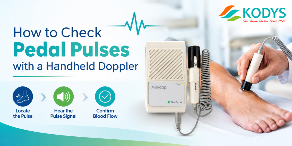
Why Use a Handheld Doppler for Pedal Pulses?
A handheld Doppler picks up blood flow in the foot that fingertips often miss. In diabetic patients, swelling, thick skin, low flow or hardened (calcified) arteries can make the dorsalis pedis and posterior tibial pulses impossible to feel. A note of “pulse absent” based on touch alone may be wrong. It may also hide early peripheral arterial disease (PAD).
A handheld Doppler turns blood flow into sound. In under five minutes per foot, a clinician can confirm whether flow is present, judge its quality from the sound, and move straight to ankle pressure and ABI. For diabetology clinics, podiatry centres, wound-care units and OPDs, it is one of the most practical tools for routine foot screening.
This guide covers the full technique, how to read the signal, the most common errors, and how to choose the right Doppler for your clinic.
What You Need
- A handheld vascular Doppler with an 8 MHz probe. 8 MHz suits shallow vessels like the foot arteries. The Minidop ES100VX is a pocket Doppler built for pedal pulses, systolic pressures and fetal heart sounds. It offers 5, 8 and 10 MHz probes and a built-in 300 mW speaker for clear sound.
- Ultrasound gel. Use plenty; air between probe and skin blocks the signal.
- A blood pressure cuff sized for the ankle, if you plan to measure ankle pressure or ABI.
- Tissue or wipes to remove gel and clean the probe after each patient.
- A quiet room, so faint signals are not lost in OPD noise.
Where to Find the Pedal Arteries
Dorsalis pedis artery (DPA): on the top of the foot, between the first and second metatarsal bones, just to the outer side of the big-toe tendon. Ask the patient to lift the big toe to make the tendon easier to see.
Posterior tibial artery (PTA): on the inner ankle, just behind and below the inner ankle bone (medial malleolus).
Peroneal artery: check it when both DPA and PTA signals are weak or absent. Its branch can be found just in front of the outer ankle bone (lateral malleolus).
Step-by-Step Technique
- Position the patient. Lay the patient flat (supine) and let them rest for 5 to 10 minutes. Remove socks, footwear and tight clothing from the legs.
- Explain the test. Tell the patient the test is painless and that they will hear a whooshing sound.
- Apply gel. Put a generous amount of gel over the artery site.
- Angle the probe. Hold the probe at 45 to 60 degrees to the skin, pointing up the leg toward the heart (against the direction of blood flow).
- Use light pressure. Press too hard and you will squeeze the artery shut and lose the signal.
- Search slowly. Move the probe in small steps and adjust the angle until you get the loudest, clearest signal.
- Listen and record. Note whether the signal is present, its type (see next section), and which artery it came from.
- Repeat on all sites, on both feet. Always compare left and right.
- Clean up. Wipe gel from the patient’s skin and clean the probe as the manufacturer recommends.
How to Interpret the Doppler Sound
| Signal | What it sounds like | What it suggests |
| Triphasic | Three distinct sounds per heartbeat, crisp and sharp | Normal, healthy artery |
| Biphasic | Two sounds per heartbeat | Mild disease, or normal in some older patients |
| Monophasic | One dull, drawn-out “whoosh” | Significant narrowing or blockage above the site |
| Absent | No arterial signal after a careful search | Severe disease or blockage; needs urgent further assessment |
| Venous | A low, windstorm-like sound that rises and falls with breathing | Vein, not artery; reposition the probe |
A monophasic or absent pedal signal should always lead to further testing: ABI, toe pressure, or referral for vascular assessment.
Common Mistakes to Avoid
- Pressing too hard. This squeezes the artery and makes the signal disappear.
- Too little gel. Air pockets block the ultrasound beam.
- Wrong probe angle. Holding the probe flat at 90 degrees gives a weak or no signal.
- Mistaking a vein for an artery. Venous sound changes with breathing; arterial sound pulses with the heartbeat.
- Rushing the search. Low-flow arteries in diabetic feet need slow, careful probe movement.
- Testing a cold foot. Cold narrows the arteries and weakens the signal. Let the patient warm up first.
- Checking only one foot. Left-right differences are often the most useful finding.
From Pulse Check to Ankle Pressure and ABI
Once you find the pedal signal, the same Doppler can measure ankle systolic pressure:
- Place the cuff just above the ankle.
- Hold the probe over the DPA or PTA.
- Inflate the cuff until the signal disappears.
- Deflate slowly. The pressure at which the signal returns is the ankle systolic pressure.
ABI = higher ankle pressure ÷ higher arm (brachial) pressure
| ABI value | Meaning |
| Above 1.40 | Non-compressible (stiff, calcified) arteries; ABI is unreliable, so check toe pressure |
| 1.00 to 1.40 | Normal |
| 0.91 to 0.99 | Borderline |
| 0.90 or below | Peripheral arterial disease |
Diabetic patients often have calcified arteries, which give falsely high ABI readings. In these cases, toe pressure and toe-brachial index (TBI) are more reliable. The Bidop ES100V3 and Smartdop 45 accept an optional PPG probe for toe pressure measurement.
For high-volume clinics, the Hadeco Smartdop XT automates ABI, TBI and pulse volume measurements with one-button operation, available in 6-port and 14-port versions.
Choosing the Right Kody Doppler for Your Clinic
| Model | Best for | Key features |
| Minidop ES100VX | GP clinics, diabetology OPDs, screening camps | Pocket size, 5/8/10 MHz bidirectional, built-in speaker, patient report software |
| Bidop ES100V3 | Clinics that need waveform records | Real-time waveform LCD, 30-waveform memory, USB, optional PPG for toe pressure |
| Smartdop 45 | Vascular and podiatry clinics needing printed reports | Waveform LCD, built-in printer, optional PPG and PVR modules |
| Bidop 7 | Advanced vascular assessment | Colour LCD, peak and mean velocity, USB data transfer, optional PPG |
| Smartdop XT (6 / 14 port) | Hospitals and high-volume diabetic foot units | Fully automatic ABI, TBI and PVR with Smart XT Link software |
As a rule of thumb: an audio-only Doppler is enough for pulse checks and manual ABI. If your clinic needs documentation, waveform records or toe pressures, choose a model with a display and PPG option.
Frequently Asked Questions
1. What frequency Doppler probe is best for pedal pulses?
An 8 MHz probe is best for pedal pulses because the dorsalis pedis and posterior tibial arteries lie close to the skin. Lower frequencies (4–5 MHz) are better for deeper vessels such as the femoral or popliteal arteries.
2. Can a handheld Doppler detect a pulse that cannot be felt?
Yes. A handheld Doppler can detect blood flow in arteries where the pulse is too weak to feel, which is common in diabetic patients with swelling, thick skin or reduced flow.
3. What does a monophasic pedal pulse signal mean?
A monophasic signal is a single dull sound per heartbeat. It usually means significant narrowing or blockage in the artery above the test site and needs further vascular assessment.
4. How do I tell an arterial signal from a venous signal?
Arterial signals pulse in time with the heartbeat. Venous signals sound like wind, are continuous, and rise and fall with breathing.
5. What angle should the Doppler probe be held at?
Hold the probe at 45 to 60 degrees to the skin, pointing toward the heart. A flat 90-degree angle gives a weak or absent signal.
6. Can the same handheld Doppler be used for ABI testing?
Yes. With a BP cuff, a handheld Doppler like the Minidop ES100VX can measure ankle systolic pressure for manual ABI calculation. For automated ABI and TBI, the Smartdop XT is designed for that workflow.
7. Why is ABI unreliable in some diabetic patients?
Diabetes can stiffen and calcify leg arteries so the cuff cannot fully compress them. This gives falsely high ABI readings above 1.40. Toe pressure or TBI is recommended in these patients.
8. How often should diabetic patients have their pedal pulses checked?
Most diabetic foot care guidelines recommend at least an annual foot examination, including pulse assessment. High-risk patients need more frequent checks as advised by their clinician.
9. Which Kody Medical Doppler is best for a small diabetic clinic?
The Minidop ES100VX suits most small clinics: it is portable, battery-powered and covers pedal pulse checks and manual ABI. Clinics needing waveform records can step up to the Bidop ES100V3.
10. Does Kody Medical provide installation and service support?
Yes. Kody Medical provides installation, training, warranty support and Annual Maintenance Contracts across India from its Chennai base. Service can be provided within 48 to 72 hours.
Equip Your Clinic for Accurate Pedal Pulse Assessment
Kody Medical Electronics supplies handheld and automatic vascular Dopplers to hospitals, diabetology clinics and podiatry centres across India. Our team can help you pick the right model for your patient volume and documentation needs, and arrange a demo.
Explore Vascular Dopplers | Call +91-9380223396 | Email kodymedical@gmail.com